Xanthidium
Desmidiaceae –
Archaeplastida
Observations on Xanthidium basiornatum
Protisten / Protisten.de – created by Wolfgang Bettighofer / Algae green-colored (2: Desmidiaceae): Xanthidium / Xanthidium basiornatum
Position in the EOL taxonomic tree. Please click on the tree view below to access EOL (Encyclopedia of Life).
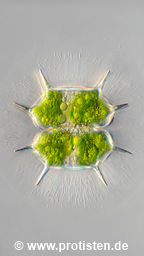

Xanthidium basiornatum (B.Eichler & Raciborski) Stastny, Skaloud & Neustupa 2013
Most likely ID: n.a.
Basionym: Xanthidium antilopaeum var. basiornatum Eichler & Raciborski 1893
Add’l Synonyms: Xanthidium canadense var. basiornatum (B.Eichler & Raciborski) Kurt Förster 1983
Sampling date 06/2026. Scale bars indicate 50 µm.
Four images. Optical journey into the cell.
In addition to a brownish elevation near the center of each half-cell, Xanthidium basiornatum is morphologically distinguished by the fact that its lateral processes, unlike those of Xanthidium antilopaeum, are not convergent or parallel, but rather distinctly divergent.
In 2013, Jan Stastny et al. demonstrated using molecular biological methods in Schwemm material that Xanthidium antilopaeum var. basiornatum is a separate species.
https://doi.org/10.1111/jpy.12051
| First: | Synoptic representation of the cell surface. The brownish elevations in the centers of the half-cells are faintly visible. |
| Second to fourth: | Optical cross-sections showing chloroplasts and oleosomes (reserve material storage). |
Please click on < or > on the image edges or on the dots at the bottom edge of the images to browse through the slides!
Place name: Moor called Schwemm near Walchsee (Tyrol, Austria)
Latitude: 47.657707 Longitude: 12.299540 Elevation: 669 m
Microscope Zeiss Universal, camera Olympus OM-D M5 MKII. DOF images.
Sampling date 06/2026. Scale bars indicate 50 µm.
Xanthidium basiornatum with chytrid parasites
Xanthidium cell, attacked by Chytridiomycota cells. The arrows point to the four chytrid cells; the rhizoids they have drilled through the protective jelly to the host cell are clearly visible (image No. 1). The cell mass of the desmid is visibly reduced (image No. 2).
The chytrid could be Rhizophydium ubiquetum, described by Hilda Canter-Lund 1961.
Canter, H. M. (1961). Studies on British chytrids. Trans. Brit. mycol. Soc, 44(2), 163-176.
| First: | Synoptic representation of the cell surface showing cell wall ornamentation. See inset with enlarged detail view top right. |
| Second: | The optical cross-section shows the remains of the chloroplasts, which, along with the rest of the cell plasma, have been eaten by the chytridiomycota. |
Please click on < or > on the image edges or on the dots at the bottom edge of the images to browse through the slides!
Place name: Moor called Schwemm near Walchsee (Tyrol, Austria)
Latitude: 47.657707 Longitude: 12.299540 Elevation: 669 m
Microscope Zeiss Axioplan, camera Olympus OM-D M5 MKII. DOF images.
© Wolfgang Bettighofer,
images under Creative Commons License V 3.0 (CC BY-NC-SA).
For permission to use of (high resolution) images please contact postmaster@protisten.de.
