Closteriaceae –
Archaeplastida
Observations on Closterium acerosum
Protisten / Protisten.de – created by Wolfgang Bettighofer / Algae green-colored (2: Desmidiaceae): Closterium / Closterium acerosum
Position in the EOL taxonomic tree. Please click on the tree view below to access EOL (Encyclopedia of Life).
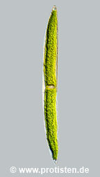

Closterium acerosum Ehrenberg ex Ralfs 1848
Most likely ID: n.a.
Basionym: n.a.
Add’l Synonyms: Vibrio acerosum Schrank 1803
Arthrodia acerosa (Ralfs) Kuntze 1891
Closterium sigmoideum Lagerheim & Nordstedt 1893
Sampling date 06/2026. Scale bars indicate 100 µm.
Four images.
| First: | Synoptic representation of the cell surface. |
| Second: | Detail view: The bars in the center mark 10 µm. The striation is very delicate and has a density of about 11 lines/10 µm. |
| Third: | Optical cross-section. |
| Fourth: | Optical cross-section showing the cell nucleus and the terminal vacuoles which contain many crystals. |
Please click on < or > on the image edges or on the dots at the bottom edge of the images to browse through the slides!
Place name: Wetland at the Pillersee (Tyrol, Austria)
Latitude: 47.531785 Longitude: 12.573095
Microscope Zeiss Universal, camera Olympus OM-D M5 MKII. DOF images.
Sampling date 06/2026. Scale bars indicate 100 µm.
Four brightfield images. Optical journey into the cell.
Please click on < or > on the image edges or on the dots at the bottom edge of the images to browse through the slides!
Place name: Wetland at the Pillersee (Tyrol, Austria)
Latitude: 47.531785 Longitude: 12.573095
Microscope Zeiss Universal, camera Olympus OM-D M5 MKII. DOF images.
Sampling date 06/2024. Scale bars indicate 100 µm.
The species is described as very slightly curved to almost straight. This specimen was very straight and could belong to Closterium acerosum f. rectum A.M. Scott & Prescott 1961. However, according to some authors, this description is invalid.
Three images.
| First: | Synoptic representation of the cell surface. The bars in the enlarged section mark 10 µm. The striation is very delicate and pale and has a density of about 11 lines/10 µm. |
| Second: | Optical cross-section showing the stelloid chloroplasts. |
| Third: | Optical cross-section showing the cell nucleus, a row of pyrenoids and the terminal vacuoles which contain many crystals. |
Please click on < or > on the image edges or on the dots at the bottom edge of the images to browse through the slides!
Place name: Wetland at the Pillersee (Tyrol, Austria)
Latitude: 47.531785 Longitude: 12.573095
Microscope Zeiss Universal, camera Olympus OM-D M5 MKII. DOF images.
© Wolfgang Bettighofer,
images under Creative Commons License V 3.0 (CC BY-NC-SA).
For permission to use of (high resolution) images please contact postmaster@protisten.de.
