Micrasterias
Desmidiaceae –
Archaeplastida
Observations on Micrasterias jenneri
Protisten / Protisten.de – created by Wolfgang Bettighofer / Algae green-colored (2: Desmidiaceae): Micrasterias / Micrasterias jenneri
Position in the EOL taxonomic tree. Please click on the tree view below to access EOL (Encyclopedia of Life).
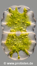

Micrasterias jenneri Ralfs 1848
Most likely ID: n.a.
Basionym: n.a.
Add’l Synonyms: Micrasterias jenneri var. simplex West 1890
Micrasterias jenneri f. brasiliensis Børgesen 1891
Micrasterias auclairii Rochou d’Aubert 1910
Sampling date 06/2025. Scale bars indicate 25 µm (1, 3–7), 5 µm (2).
Seven images. Optical journey into the cell.
| First: | Synoptic representation of the cell wall structure. The granules visible on the surface do not represent cell wall structuring, but are patches of the mucilaginous sheath. |
| Second: | Zoom-in showing the tiny cell wall pores. The gelatinous mass enveloping the cell is transported outwards through these pores. |
| Third to seventh: | Optical cross-section showing parts of the chloroplasts and several circular, luminous structures, spherical assimilate bodies with storage lipids (oleosomes). |
Please click on < or > on the image edges or on the dots at the bottom edge of the images to browse through the slides!
Place name: Moor called Schwemm near Walchsee (Tyrol, Austria)
Latitude: 47.657707 Longitude: 12.299540 Elevation: 669 m
Microscope Zeiss Axioplan, camera Olympus OM-D M5 MKII. DOF images.
Sampling date 06/2025. Scale bars indicate 50 µm.
Four images.
| First: | Synoptic representation of parts of the cell wall structure. The granules visible on the surface do not represent cell wall structuring, but are patches of the mucilaginous sheath. |
| Second to fourth: | Optical cross-section showing chloroplasts, the central nucleus with nucleoli and several circular, luminous structures, spherical assimilate bodies with storage lipids (oleosomes). |
Please click on < or > on the image edges or on the dots at the bottom edge of the images to browse through the slides!
Place name: Moor called Schwemm near Walchsee (Tyrol, Austria)
Latitude: 47.657707 Longitude: 12.299540 Elevation: 669 m
Microscope Zeiss Axioplan, camera Olympus OM-D M5 MKII. DOF images.
Sampling date 06/2025. Scale bars indicate 50 µm.
Three brightfield images.
| First: | Synoptic representation of parts of the cell wall structure. The granules visible on the surface do not represent cell wall structuring, but are patches of the mucilaginous sheath. |
| Second and third: | Optical cross-section showing chloroplasts, the central nucleus with nucleoli and several circular, luminous structures, spherical assimilate bodies with storage lipids (oleosomes). |
Please click on < or > on the image edges or on the dots at the bottom edge of the images to browse through the slides!
Place name: Moor called Schwemm near Walchsee (Tyrol, Austria)
Latitude: 47.657707 Longitude: 12.299540 Elevation: 669 m
Microscope Zeiss Axioplan, camera Olympus OM-D M5 MKII. DOF images.
© Wolfgang Bettighofer,
images under Creative Commons License V 3.0 (CC BY-NC-SA).
For permission to use of (high resolution) images please contact postmaster@protisten.de.
