Micrasterias
Desmidiaceae –
Archaeplastida
Observations on Micrasterias rotata
Protisten / Protisten.de – created by Wolfgang Bettighofer / Algae green-colored (2: Desmidiaceae): Micrasterias / Micrasterias rotata
Position in the EOL taxonomic tree. Please click on the tree view below to access EOL (Encyclopedia of Life).


Micrasterias rotata Ralfs 1848
Most likely ID: n.a.
Basionym: n.a.
Add’l Synonyms: Echinella rotata Greville 1833
Cosmarium stellinum Corda 1835
Eutomia rotata (Greville) Harvey 1841
Micrasterias rotata f. granulata West 1892
Micrasterias rotata f. evoluta W.B.Turner 1893
Micrasterias rotata var. pulchra Lemmermann 1896
Micrasterias rotata var. evoluta (W.B.Turner) Willi Krieger 1939

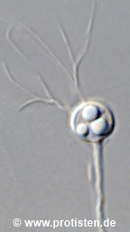
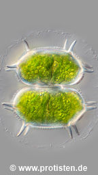
Sampling date 04/2007. Scale bars indicate 50 µm.
Three images, two of them in a slide changer.
| First: | Synoptic representation of the cell surface. The image shows a very special view of Micrasterias rotata using an inverted microscope. The cell stands vertically on the slide. Like most desmids, Micrasterias can move by secreting mucus; they usually move toward light. In the dark, the cells stand upright. |
| Second: | Synoptic representation of the cell surface. |
| Third: | Optical cross-section through the cell showing chloroplasts with numerous pyrenoids and nucleus with its papillose nucleolus. |
Please click on < or > on the image edges or on the dots at the bottom edge of the images to browse through the slides!
Place name: Bog near Salzburg (Austria)
Latitude: 48.068516 Longitude: 12.954134
(1): Microscope Zeiss IM35, camera Olympus C7070WZ. DOF image.
(2, 3): Microscope Zeiss Universal, camera Olympus C7070WZ. DOF images.
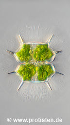
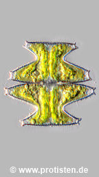
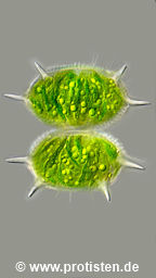
Sampling date 04/2021. Scale bars indicate 50 µm (3, 4), 100 µm (5).
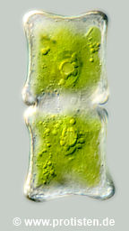
Five images.
| First and second: | Overview using stereo microscope. |
| Third: | Synoptic representation of the chloroplasts surface structure. |
| Fourth: | Optical cross-section through the cell showing chloroplasts with pyrenoids and the nucleus. |
| Fifth: | Cell division in its final phase. |
Please click on < or > on the image edges or on the dots at the bottom edge of the images to browse through the slides!
Place name: Pond near Großostheim (Germany)
Latitude: 49.88482168 Longitude: 9.09980822
(1, 2): Stereo microscope Olympus SZX16, camera Olympus OM-D M5 MKII.
(3, 4): Microscope Zeiss Axioplan, camera Olympus OM-D M5 MKII. DOF images.
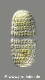
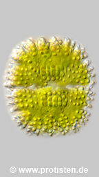
Sampling date 06/2023. Scale bars indicate 50 µm.
Three images.
| First and second: | Optical cross sections through the chloroplasts showing numerous oleosomes, circular, luminous structures, which are spherical assimilate bodies with storage lipids. |
| Second: | Synoptic representation of the cell surface. |
| Third: | Optical cross-section through the cell showing chloroplasts with numerous pyrenoids and nucleus with its papillose nucleolus. |
Please click on < or > on the image edges or on the dots at the bottom edge of the images to browse through the slides!
Place name: Wetland Lauchseemoor, Fieberbrunn (Tyrol, Austria)
Latitude: 47.46954439 Longitude: 12.53826499 Elevation: 866 m
Microscope Zeiss Axioplan, camera Olympus OM-D M5 MKII. DOF images.
© Wolfgang Bettighofer,
images under Creative Commons License V 3.0 (CC BY-NC-SA).
For permission to use of (high resolution) images please contact postmaster@protisten.de.

New items
New items
ubiquetum
basiornatum
pinnatifida
elegantissimiforme
antilopaeum
laeve
caelatum
decedens