


elegantissimiforme
pseudowillei
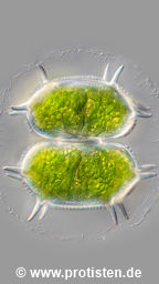
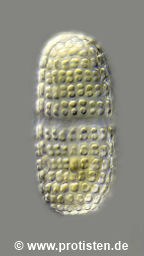
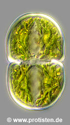
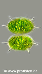
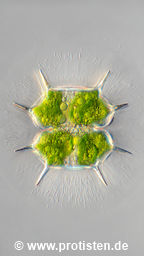
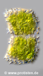
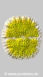
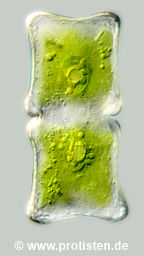
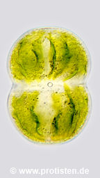
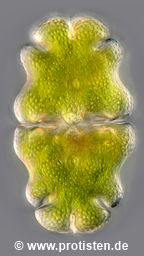
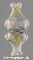
antilopaeum
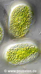
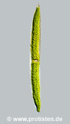
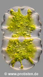
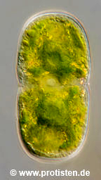
laeve
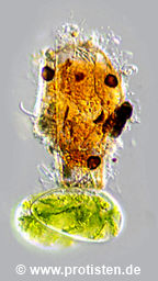
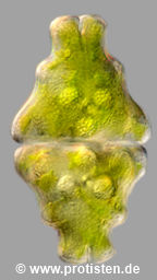
basiornatum
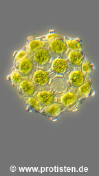
capitulum
spec.
africana
caelatum
pinnatifida
striolatum
decedens

acutiformis
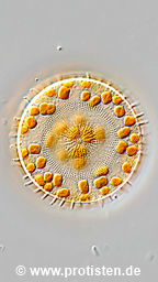
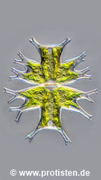
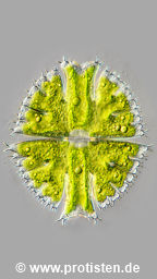
astraea
bipyramidatum
cinctum
pulchrum
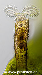
melicerta
lunatus
brebissonii
montanum
directum
truncatum
vestitum
var. vestitum
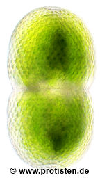
meneghinianum
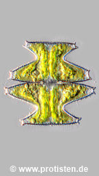
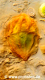
didelta
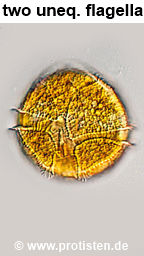
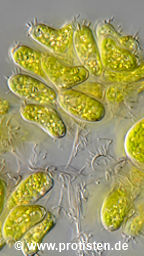
furcata
jenneri
ventricosum
insigne
cucurbita
fimbriata
cucurbita
wailesii
filifera
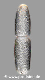
brightwellii
elegantissimiforme
pseudowillei
antilopaeum
laeve
basiornatum
capitulum
spec.
africana
caelatum
pinnatifida
striolatum
decedens
acutiformis
astraea
bipyramidatum
cinctum
pulchrum
melicerta
lunatus
brebissonii
montanum
directum
truncatum
vestitum
var. vestitum
meneghinianum
didelta
furcata
jenneri
ventricosum
insigne
cucurbita
fimbriata
cucurbita
wailesii
filifera
brightwellii