Observations on Ditylum brightwellii
Protisten / Protisten.de – created by Wolfgang Bettighofer / Bacillariophyta / Mediophyceae / Lithodesmiales / Ditylum brightwellii
Position in the EOL taxonomic tree. Please click on the tree view below to access EOL (Encyclopedia of Life).


Ditylum brightwellii (T.West) Grunow 1885
Most likely ID: n.a.
Basionym: Triceratium brightwellii T.West 1860
Add’l Synonyms: Ditylum trigonum L.W.Bailey ex L.W.Bailey 1862
Ditylum inaequale Bailey ex L.W.Bailey 1862
Sampling date 01/2022. Scale bars indicate 100 µm.
Two images.
Please click on < or > on the image edges or on the dots at the bottom edge of the images to browse through the slides!
Place name: Baltic Sea, Kieler Förde, Kiel Fjord (Germany)
Latitude: 54.3894126 Longitude: 10.1749055
Stereo microscope Olympus SZX16/Planapo 2.0x, camera Olympus OM-D M5 MKII. DOF images.
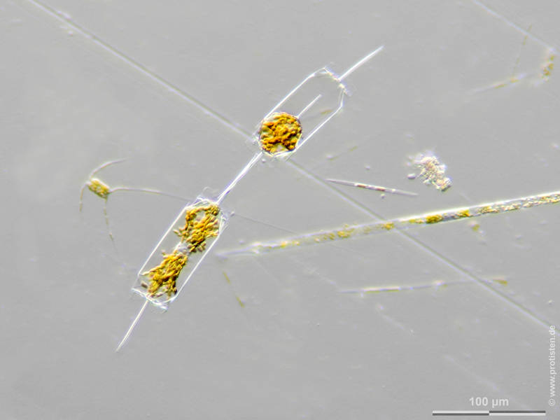
Sampling date 01/2022. Scale bar indicates 100 µm.
The cell in the upper part has developed into a resting cyst.
Koester, J. A., Brawley, S. H., Karp-Boss, L., & Mann, D. G. (2007). Sexual reproduction in the marine centric diatom Ditylum brightwellii (Bacillariophyta). European Journal of Phycology, 42(4), 351-366. https://doi.org/10.1080/09670260701562100
Please click on < or > on the image edges or on the dots at the bottom edge of the images to browse through the slides!
Place name: Baltic Sea, Kieler Förde, Kiel Fjord (Germany)
Latitude: 54.3894126 Longitude: 10.1749055
Stereo microscope Olympus SZX16/Planapo 2.0x, camera Olympus OM-D M5 MKII. DOF image.
Sampling date 12/2025. Scale bars indicate 50 µm (1–3), 10 µm (4–6).
Four images. Two cells starting to produce a resting cyst.
| First to third: | Synoptic representation of the complete frustule and optical cross sections. |
| Fourth to sixth: | Enlarged sections showing details of the frustules. |
Please click on < or > on the image edges or on the dots at the bottom edge of the images to browse through the slides!
Place name: Baltic Sea, Kieler Förde, Kiel Fjord (Germany)
Latitude: 54.3894126 Longitude: 10.1749055
Microscope Zeiss Axioplan, camera Olympus OM-D M5 MKII. DOF images.
Sampling date 12/2025. Scale bars indicate 50 µm (1), 10 µm (3).
Three images.
| First: | Complete frustule. |
| Second and third: | Close-up views of the valves. Interestingly, the valve bristles can have different structures within a single cell, ranging from needle-shaped to paddle-shaped. |
Please click on < or > on the image edges or on the dots at the bottom edge of the images to browse through the slides!
Place name: Baltic Sea, Kieler Förde, Kiel Fjord (Germany)
Latitude: 54.3894126 Longitude: 10.1749055
Tescan Clara SEM, © Wolfgang Bettighofer/Jürgen Stampfl.
Sampling date 12/2025. Scale bars indicate 2 µm (1), 1 µm (2), 200 nm (3).
Three images.
| First: | This close-up shows the seam between the valve and the girdle band. The hole types of the valve and girdle band are different. |
| Second and third: | Detailed images of the C-shaped openings through the valve. |
Please click on < or > on the image edges or on the dots at the bottom edge of the images to browse through the slides!
Place name: Baltic Sea, Kieler Förde, Kiel Fjord (Germany)
Latitude: 54.3894126 Longitude: 10.1749055
Tescan Clara SEM, © Wolfgang Bettighofer/Jürgen Stampfl.
Sampling date 12/2025. Scale bars indicate 10 µm (1), 2 µm, (2), 1 µm (3), 2 µm (4).
Four images.
| First: | A triangular-shaped Valve (Ditylum brightwellii forma trigonum) with shovel-shaped projections. |
| Second and third: | Detailed images of the C-shaped openings through the valve. |
| Fourth: | Close-up of a small part of the shovel-shaped brush ring. |
Please click on < or > on the image edges or on the dots at the bottom edge of the images to browse through the slides!
Place name: Baltic Sea, Kieler Förde, Kiel Fjord (Germany)
Latitude: 54.3894126 Longitude: 10.1749055
Tescan Clara SEM, © Wolfgang Bettighofer/Jürgen Stampfl.
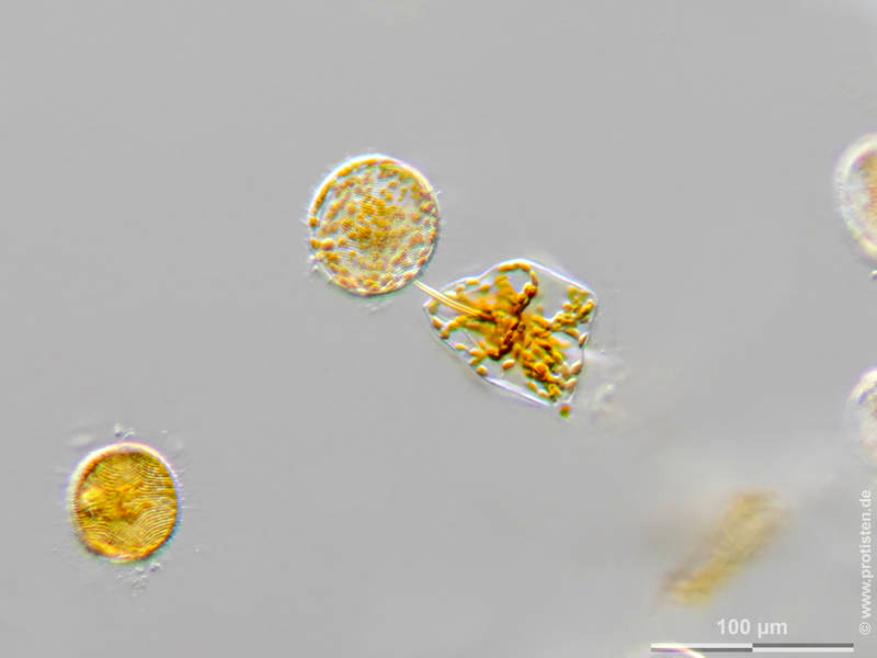
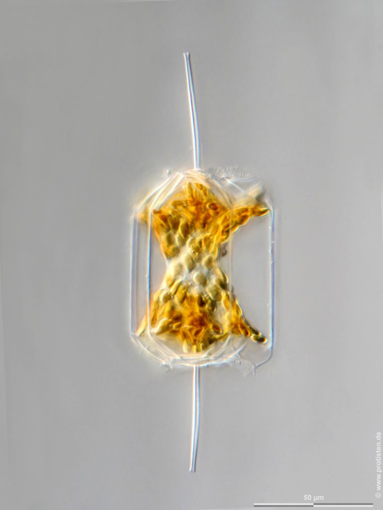
Sampling date 12/2025. Scale bars indicate 100 µm (1), 50 µm (2).
Two images of Ditylum brightwellii forma trigonum.
| First: | View into a Petri dish through a stereomicroscope. |
| Second: | The same cell under the microscope. Planapo 20*/0.8, DIC, DOF image. |
Please click on < or > on the image edges or on the dots at the bottom edge of the images to browse through the slides!
Place name: Baltic Sea, Kieler Förde, Kiel Fjord (Germany)
Latitude: 54.3894126 Longitude: 10.1749055
(1) Stereo microscope Olympus SZX16/Planapo 2.0x, camera Olympus OM-D M5 MKII. DOF image.
(2) Microscope Zeiss Axioplan, camera Olympus OM-D M5 MKII. DOF image.
Sampling date 04/2017. Scale bars indicate 50 µm.
Four images.
| First: | Synoptic representation of the frustule. |
| Second: | The same cell in cross section. |
| Third: | Cell shortly after a binary fission. |
| Fourth: | Two mature cells, resulting from cell division, are connected at their labiate processes by a gelatinous junction. |
Please click on < or > on the image edges or on the dots at the bottom edge of the images to browse through the slides!
Place name: North Sea around Heligoland
Latitude: 54.186311 Longitude: 7.895034
Microscope Zeiss Universal, camera Olympus C7070WZ. DOF images.
© Wolfgang Bettighofer,
images under Creative Commons License V 3.0 (CC BY-NC-SA).
For permission to use of (high resolution) images please contact postmaster@protisten.de.
