Observations on Coscinodiscus concinnus
Protisten / Protisten.de – created by Wolfgang Bettighofer / Bacillariophyta / Coscinodiscophyceae / Coscinodiscales / Coscinodiscus concinnus
Position in the EOL taxonomic tree. Please click on the tree view below to access EOL (Encyclopedia of Life).


Coscinodiscus concinnus W.Smith 1856
Most likely ID: n.a.
Basionym: n.a.
Add’l Synonyms: n.a.
Sampling date 04/2017.
Three images.
Please click on < or > on the image edges or on the dots at the bottom edge of the images to browse through the slides!
Place name: North Sea around Heligoland
Latitude: 54.186311 Longitude: 7.895034 Elevation: 0 m
Leica dissecting microscope, camera Olympus OM-D E-M5 II. DOF images.

Sampling date 01/2022. Scale bars indicate 100 µm.
Five images, four of them in a slide changer.
Please click on < or > on the image edges or on the dots at the bottom edge of the images to browse through the slides!
Place name: Baltic Sea, Kieler Förde, Kiel Fjord (Germany)
Latitude: 54.3894126 Longitude: 10.1749055 Elevation: 0 m
Stereo microscope Olympus SZX16/Planapo 2.0x, camera Olympus OM-D E-M5 II. DOF images.
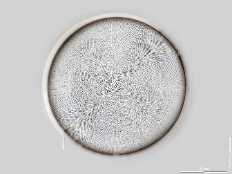
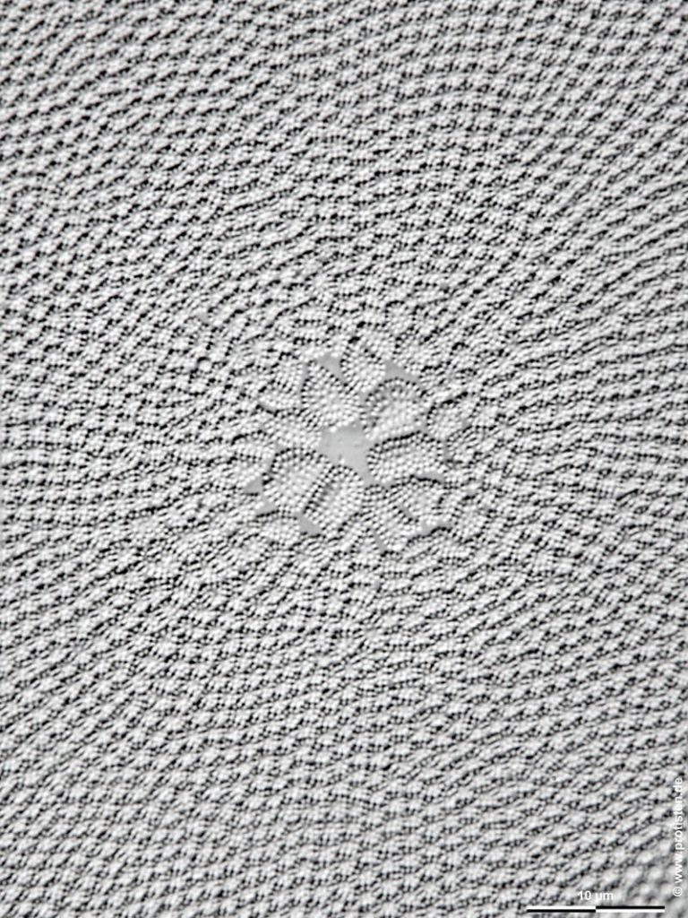
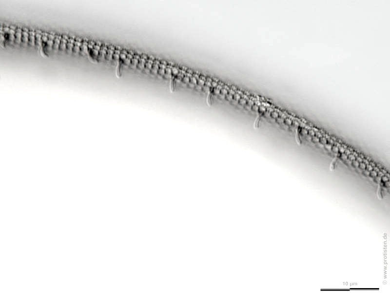
Sampling date 04/2025. Scale bars indicate 100 µm (1), 10 µm (2, 3).
Sampling and preparation for the light microscope by Hermann Hochmeier, Wilhelmsburg (Austria).
Three images.
| First: | Synoptic representation of the valve. |
| Second: | Detail of the valve center showing the fine sieve pores (called cribra) of the outer surface of the frustule. |
| Third: | Internal view of the valve margin. The processes are called rimoportulae or labiate processes; they secrete mucilage that serves to adhere to a substrate. This phenomenon is shown here. |
Place name: Heligoland harbor
Latitude: 54.177119 Longitude: 7.893548 Elevation: 0 m
Microscope Zeiss Axioplan, camera Olympus OM-D M5 MKII. DOF images.
Sampling date 04/2025. Scale bars indicate 100 µm (1), 5 µm (2), 1 µm (3).
Sampling and preparation for the light microscope by Hermann Hochmeier, Wilhelmsburg (Austria).
Three images.
| First: | Valve, inner surface. |
| Second and third: | A high-magnification view of the inner girdleband surface reveals its double-layered structure. The delicate sieve areas (cribra) on the outer surface are visible through the larger openings of the inner perforated plate. |
Please click on < or > on the image edges or on the dots at the bottom edge of the images to browse through the slides!
Place name: Heligoland harbor
Latitude: 54.177119 Longitude: 7.893548 Elevation: 0 m
Tescan Clara SEM, © Wolfgang Bettighofer/Jürgen Stampfl
Sampling date 04/2025. Scale bars indicate 20 µm (1), 5 µm (2), 2 µm (3).
Sampling and preparation for the light microscope by Hermann Hochmeier, Wilhelmsburg (Austria).
Three images.
| First: | At medium magnification, the row of rimoportulae (labiate processes) can already be seen on the inner edge of the valve. |
| Second and third: | Labiate processes at high magnification. |
Please click on < or > on the image edges or on the dots at the bottom edge of the images to browse through the slides!
Place name: Heligoland harbor
Latitude: 54.177119 Longitude: 7.893548 Elevation: 0 m
Tescan Clara SEM, Tescan Clara SEM, © Wolfgang Bettighofer/Jürgen Stampfl
© Wolfgang Bettighofer,
images under Creative Commons License V 3.0 (CC BY-NC-SA).
For permission to use of (high resolution) images please contact postmaster@protisten.de.
