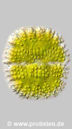
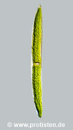
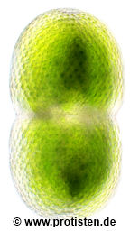
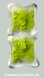
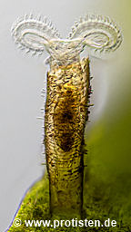
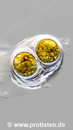
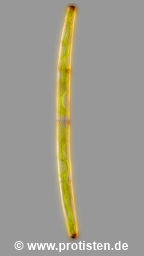
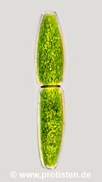
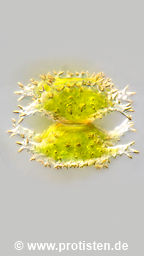
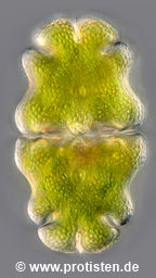
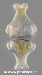
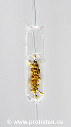
Platycola Kent, 1882
Most likely ID: Platycola decumbens (Ehrenberg, 1830) Kent, 1882
Basionym: Vaginicola decumbens Ehrenberg, 1830
Add’l Synonyms: Vaginicola truncata Fromentel, 1876
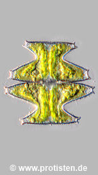
Note: According to Kahl (1935) and Foissner (1992), the members of the Platycola species can be distinguished by their shell shapes. In contrast, Warren (1983) writes that Platycola decumbens can only be distinguished from P. dilatata by the number of contractile vacuoles (CVs). The shapes of the shells should be variable and couldn’t be used as a distinguishing feature. He argues that de Fromentel, when first describing P. dilatata in 1874, described the presence of two CVs.
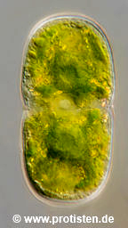
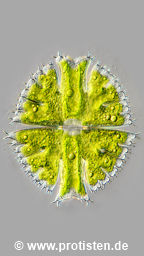
I consider this to be a misinterpretation in the original description. All other peritrichs have only one CV, and they are always located near the peristome. Even very large cells, such as those in Thuricola folliculata, require only one CV!
If you squeeze the cells too hard during microscopic observation, it may look as if they have more than one CV! This is how the description with 2 CVs came about.
In his plate X, de Fromentel also depicted two contractile vacuoles in Cothurnia elongata (Fig. 14, now Vaginicola elongata). Kahl notes that this must be a misinterpretation!
de Fromentel, E., & Jobard-Muteau, J. (1874). Etudes sur les microzoaires, ou infusoires proprement dits: comprenant de nouvelles recherches sur leur organisation, leur classification et la description des espèces nouvelles ou peu connues. G. Masson.
Kahl, A. (1935). Urtiere oder Protozoa. I. Wimpertiere oder Ciliata (Infusoria). 4. Peritricha und Chonotricha. Die Tierwelt Der Angrenzenden Meeresteile, 30, 651-886.
Warren, A. (1983). The ecology, morphology and taxonomy of freshwater peritrich ciliates. University of Surrey (United Kingdom).
(Thesis submitted for the degree of Doctor of Philosophy in the University of Surrey)
Foissner, W. , Berger, H., Kohmann, F. (1992). Taxonomische und ökologische Revision der Ciliaten des Saprobiensystems: Band II: Peritrichia, Heterotrichida, Odontostomatida (Vol. 5/92). Bayerisches Landesamt für Wasserwirtschaft.